. 2024 Oct 24;6(5):e240041. doi: 10.1148/ryct.240041

Figure 4:

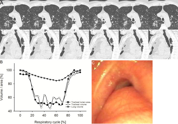
Representative example of circumferential-type tracheobronchomalacia (TBM). (A) Image from four-dimensional (4D) CT (noncontrast, lung window) in a 72-year-old male patient with circumferential-type TBM at the level of maximum observed tracheal collapsibility at representative selected time points of the respiratory cycle. The upper row shows axial view, the lower row sagittal view. The axial view demonstrates the circular collapse of the trachea because of cartilaginous malacia. (B) Graph shows the changes of minimal tracheal lumen area, tracheal volume, and lung volume in the same patient over the respiratory cycle with 21 time points in 5%-wide steps at 4D CT. (C) Image from correlated bronchoscopy.

Representative example of circumferential-type tracheobronchomalacia (TBM). (A) Image from four-dimensional (4D) CT (noncontrast, lung window) in a 72-year-old male patient with circumferential-type TBM at the level of maximum observed tracheal collapsibility at representative selected time points of the respiratory cycle. The upper row shows axial view, the lower row sagittal view. The axial view demonstrates the circular collapse of the trachea because of cartilaginous malacia. (B) Graph shows the changes of minimal tracheal lumen area, tracheal volume, and lung volume in the same patient over the respiratory cycle with 21 time points in 5%-wide steps at 4D CT. (C) Image from correlated bronchoscopy.