. 2024 Nov 6;12:RP89755. doi: 10.7554/eLife.89755

Figure 5. Mutation of GluK1-1a splice insert residues affects the receptor modulation by Neto proteins.

Bar graphs (mean ± SEM) show a comparison between wild-type and mutant receptors with Neto proteins for different kinetic properties. (A) Mean-weighted Tau (τDes) values for GluK1-1a wild-type and mutant receptors in the presence of 10 mM glutamate and expressed with Neto1/2. (B) Tau (τRecovery) recovery values for GluK1-1a and mutants with Neto1/2. (C) The ratio of the peak amplitudes evoked in the presence of 1 mM kainate and 10 mM glutamate for GluK1-1a mutants co-expressed with Neto1/2 is shown. (D) The rectification index represented by the ratio of currents evoked by 10 mM glutamate application at +90 mV and –90 mV for the wild-type and mutant receptors with Neto proteins is shown. The wild-type GluK1 splice variants’ data is the same as in Figure 2 and is replotted here for comparison. Error bars indicate mean ± SEM, N in each bar represents the number of cells used for analysis, and * indicates the significance at a 95% confidence interval.

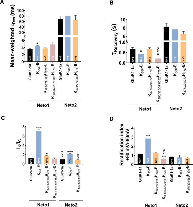
Figure 5—source data 1. Data used for the electrophysiology plots.

Figure 5.

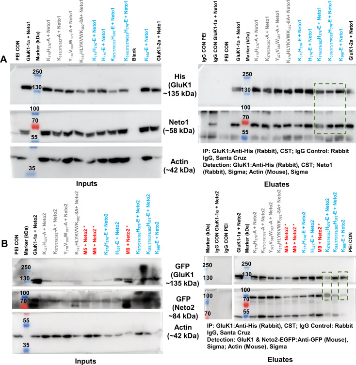
Figure 5—figure supplement 1. Co-immunoprecipitation analysis of GluK1-1a splice mutants and Neto proteins.

Figure 5—figure supplement 1.

(A, B) represent the raw western blots for receptor pull-down experiments using His-antibody (CST). The left and right panels show the inputs and eluates for the co-IP. Actin was used as an internal control. The antibodies used to detect receptor or Neto proteins have been indicated. Rabbit IgG controls were set up using WT receptor with Neto1 or Neto2 and negative control along with the test samples to check for non-specific interactions. All the experiments were done in triplicates. Charge-neutral (Ala) mutants are labeled in gray, charge reversal (Glu) mutants in cyan and marker, and controls in black. Red asterisks are mutants not used for further studies. The molecular weight of markers and the expected proteins have been indicated. Green boxes display that the mutant receptors shown in the functional analysis were able to interact with both Neto proteins efficiently when co-transfected with Neto1 or Neto2. The western blots are raw images without any editing, they are assembled as they were cut from the same blot for probing with respective primary and secondary antibodies as mentioned in the panels A and B.
Figure 5—figure supplement 1—source data 1. PDF files containing labelled western blots for Figure 5—figure supplement 1A.
Figure 5—figure supplement 1—source data 2. Original files for western blots displayed in Figure 5—figure supplement 1A.
Figure 5—figure supplement 1—source data 3. PDF files containing labelled western blots for Figure 5—figure supplement 1B.
Figure 5—figure supplement 1—source data 4. Original files for western blots displayed in Figure 5—figure supplement 1B.
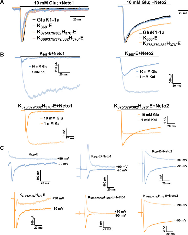
Figure 5—figure supplement 2. Representative traces for the GluK1 mutant receptors with Neto proteins.

Figure 5—figure supplement 2.

(A) Demonstrates the normalized traces for wild-type and mutant receptors with Neto1/2 for glutamate-evoked desensitization. (B) Displays representative traces for glutamate vs. kainate evoked responses in the presence of Neto1 or Neto2. (C) Representative 100 ms traces for currents evoked at positive (+90 mV) and negative (–90 mV) potentials in the absence and presence of Neto proteins.
Figure 5—figure supplement 2—source data 1. Data used for the electrophysiology plots.