Figure 2.

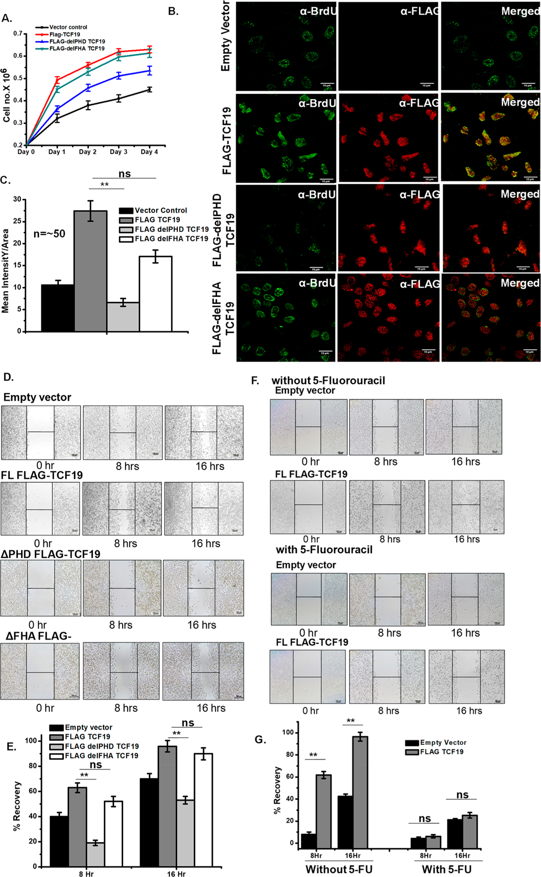
TCF19 promotes cellular proliferation. (A) Cell proliferation assay by the cell number counting technique. The WT-TCF19 overexpressed cell (red) proliferates at a rate higher than those of delPHD-TCF19 (blue) overexpressed and control (black) cells. (B) BrdU incorporation assay by confocal microscopy measurement also showed higher proliferation in WT-TCF19 overexpressed cells. (C) Quantification of the microscopy data (n ∼ 50). (D) The wound healing assay also showed a positive result on proliferation in the case of wild type overexpressed cells. (E) Quantification of recovery by wound healing. (F) Wound healing assay in the presence and absence of potential proliferation blocker 5-fluorouracil in empty vector transfected cells as well as WT-TCF19 transfected cells. (G) Quantification of wound healing in the presence and absence of 5-fluorouracil.
