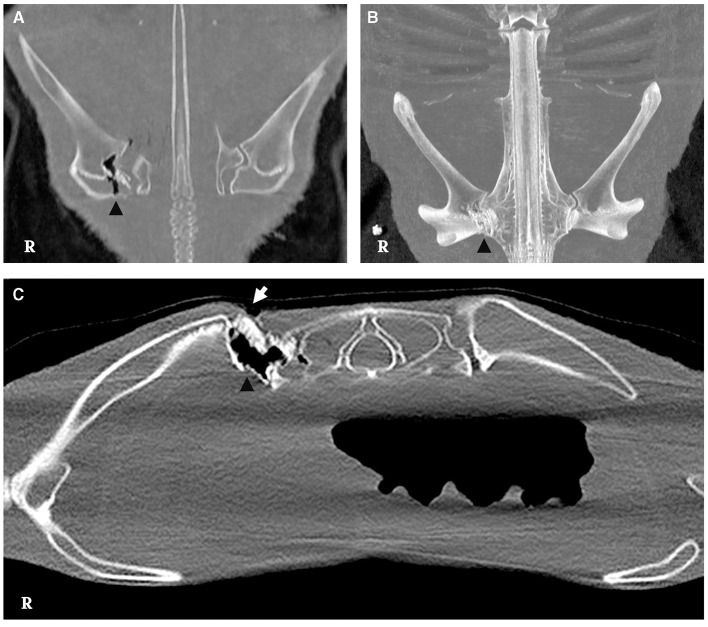
Figure 3.
CT scan of the lesion. (A, B) Irregular borders are observed in the medial aspect of the scapulocoracoid cartilage with presence of slight gas attenuation (arrowhead) [bone window, dorsal multiplanar reconstruction (MPR) and dorsal maximum intensity projection 3D (MIP), respectively]. (C) Bone window, cross-sectional image, displaying discontinuity of the skin on the dorsal aspect of the scapulocoracoid-synarcual joint (white arrow), featuring irregular margins and gas attenuation presence (arrowhead).

