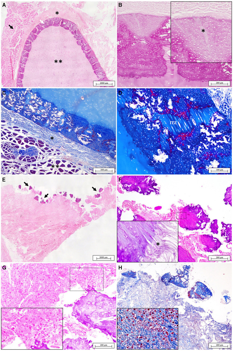
Figure 6.
(A) Normal structure of healthy cartilage displaying four distinct layers, from the outer to the inner: the muscle layer (black arrow), the perichondrium (*), tesserae layer (white arrow) and the unmineralized cartilage core (**) (H&E). (B) Normal tesserae. Sharpey's fibers from the perichondrium are observed penetrating in the cap zone of the tesserae (*) (PAS). (C) Normal structure of the healthy cartilage stained with MT. The muscle layer is evident in red (**) while the perichondrium (*) and the mineralized tesserae layer (white arrow) are seen in intense blue (MT). (D) A detail of the fibrous intertesserae zone (ITZ) stained in blue with MT. The edges of each tesserae piece are shown marked in red probably due to different types and degrees of maturation of collagen fibers (MT). (E) Lesion in the scapulocoracoid cartilage of the affected animal. The injured area is characterized by the disorganization of the tesserae layer (arrows), with the presence of broken tesserae pieces over an abundant fibrous connective tissue (H&E). (F) Detailed of fractured pieces of tesserae displaced from the adjacent perichondrium (H&E). (Inset) fraying of the Sharpey's fibers were observed in each piece of tesserae (*) (PAS). (G) The fibrous tissue below the broken tesserae layer appears increased and infiltrated by an abundant inflammatory cell population (H&E). (H) The MT stain revealed the absence of the muscle layer while it helps to identify granulocytes as the main cells of the inflammatory infiltrate (MT).

