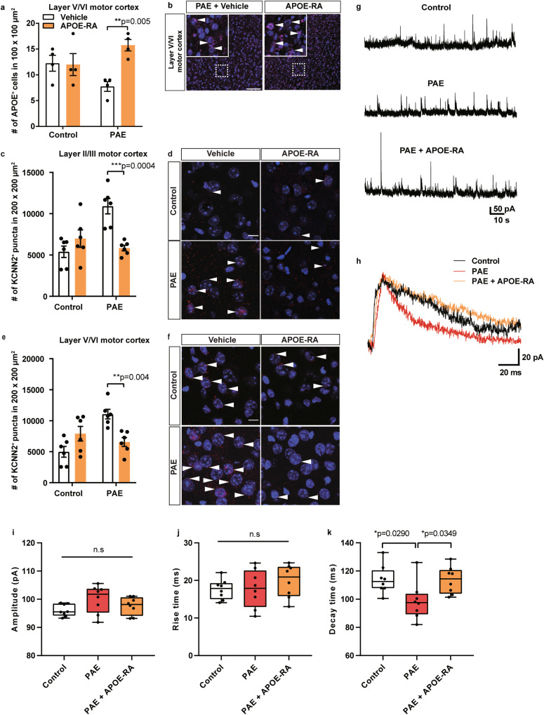
Fig. 3. Postnatal APOE-RA treatment alleviated the decreased endogenous APOE level, excessive KCNN2, and shorter decay time of NMDAR-mediated sEPSC in the motor cortex in PAE mice.
a APOE-RA treatment increases the number of APOE-positive cells that is reduced by PAE in layer V/VI of the motor cortex (n = 4 per group). **P < 0.01 by two-way ANOVA with simple main effect test. Data represent mean ± s.e.m. b Representative images of APOE (magenta, arrowheads) and DAPI (blue) staining in layer V/VI of the motor cortex in PAE mice treated with vehicle or APOE-RA. Dotted boxes indicate the region shown at higher magnification in the inset. The number of KCNN2 puncta is significantly decreased in layer II/III (c) and layer V/VI of the motor cortex (e) in PAE mice after the APOE-RA treatment compared to vehicle treatment (n = 6 per group). **P < 0.01, ***P < 0.001 by two-way ANOVA with simple main effect test. Data represent mean ± s.e.m. d, f Representative images of KCNN2 (red, arrowheads) and DAPI (blue) staining. Scale bars = 10 µm. g Examples of NMDAR-mediated sEPSCs recorded under the voltage-clamp condition at a holding potential of +50 mV in pyramidal neurons of layer V/VI in the indicated experimental groups. h NMDAR-mediated sEPSCs averaged from 5 events in control, PAE, and PAE + APOE-RA mice. i–k No significant differences are observed in the amplitude (i) and rise time (j) between the groups. PAE group shows a significantly shorter decay time than control and APOE-RA treated PAE group (k). Box plot represents the 25th, median, and 75th percentile. n = 8 cells per group. Whiskers extend to min and max values. *P < 0.05 by One-way ANOVA with Tukey’s post hoc test.

