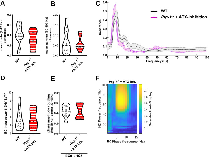
Fig. 4. ATX inhibition restores entorhinal-hippocampal coherence and PAC to control values.
A Inhibition of the LPA-synthesizing molecule ATX by PF8380 (ATX inh.) increased EC-HC theta coherence (7–12 Hz) in Prg-1−/− mice to WT levels (n = 15 WT mice, 7 Prg-1−/− + ATX inh. mice, Bayesian analysis). B Low and high gamma coherence (30–100 Hz) in the entorhinal-hippocampal network in Prg-1−/− mice was reduced to WT levels following ATX inhibition (n = 15 WT mice and 7 Prg-1−/− + ATX inh. mice, Bayesian analysis). C Coherence spectrum of WT and Prg-1−/− mice following ATX-inhibition shows restored coherence to WT levels (n = 15 WT mice, 7 Prg-1−/− + ATX inh. mice, Bayesian analysis). D EC theta power (10 Hz) in Prg-1−/− animals was increased to WT levels following ATX-inhibition by PF8380 (n = 14 WT, 7 Prg-1−/− + PF8380, Bayesian analysis). E PAC of EC theta oscillation (10 Hz, EC θ) and hippocampal gamma power (30–100 Hz, EC γ) in Prg-1−/− mice increased back to wild type levels following inhibition of the LPA-synthesizing enzyme ATX (ATX inh.) (n = 15 wild type mice, 7 Prg-1−/− + ATX inh. mice, Bayesian analysis). F Representative PAC of a Prg-1−/− animal following ATX-inhibition. Note the restored PAC between the entorhinal theta frequency (10 Hz) and the hippocampal gamma power (30–100 Hz) in the Prg-1−/− animal after ATX inhibition. A, B, D, E Data are represented as violin plots covering all individual data points. Median, lower and upper quartiles are shown by dotted linie (* and ** show group differences of * >80% or **>90% for Bayesian analysis).

