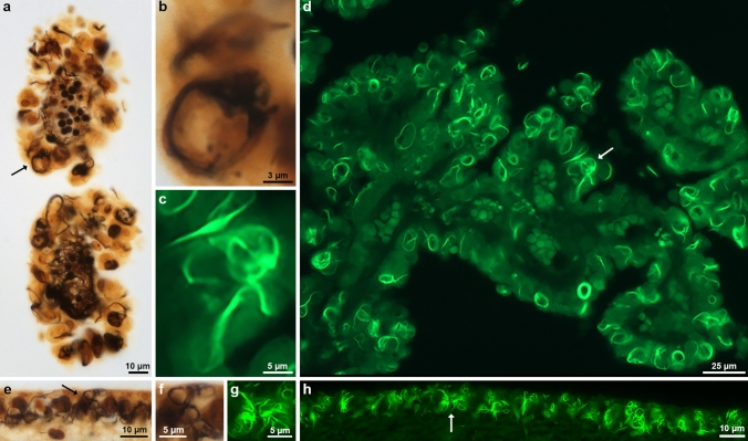
Fig. 1.
Histopathology of Biondi bodies using Bielschowsky silver and Thioflavin S. Choroid plexuses (a,d) and ependymal linings of the lateral ventricle (e,h) from cases 1 and 3 were stained with modified Bielschowsky silver (a,b,e,f) and Thioflavin S (c,d,g,h). Biondi bodies are seen in the cytoplasm of ependymal cells as argyrophilic (a,b,e,f) or fluorescent (c,d,g,h) inclusions. Arrows (a,d,e,h) point to Biondi bodies seen at high power in b, c, f and g. Scale bars: 10 µm (a,e,h), 3 µm (b), 5 µm (c,f,g), and 25 µm (d)

