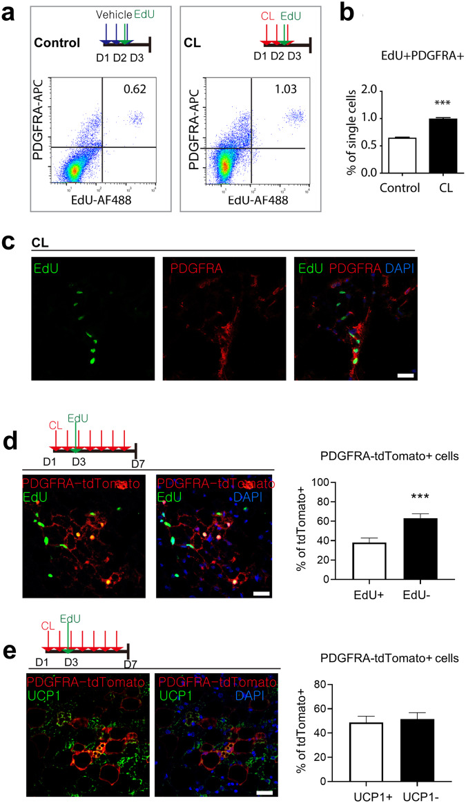
Fig. 1. Proliferation and de novo differentiation of PDGFRA+ progenitors after CL stimulation.
a Flow cytometric analysis of PDGFRA expression and EdU incorporation. b Quantification of the percentages of EdU+ PDGFRA+ cells under control and CL treatment conditions. c Images of EdU and PDGFRA staining in paraffin sections of iWAT from mice treated with CL for 3 days and injected with EdU 4 h before analysis. Bar = 25 μm. d Immunostaining of EdU and tdTomato in paraffin sections of iWAT from PDGFRA-CreER/tdTomato reporter mice treated with CL for 7 days and injected with EdU on Day 3. Bar = 25 μm. e Immunostaining of UCP1 and tdTomato in paraffin sections of iWAT from PDGFRA-CreER/tdTomato reporter mice treated with CL for 7 days and injected with EdU on Day 3. Bar = 25 μm. The data were analyzed by an unpaired two-tailed t test (mean ± SEM, n = 5–8; ***P < 0.001).

