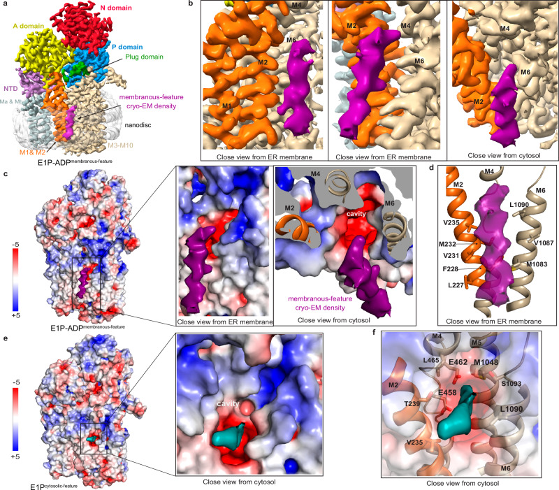
Fig. 4. The cytosol-open E1P-ADP and E1P states with associated cryo-EM density features.
a 3.4 Å global resolution cryo-EM map of the E1P-ADP state, colored as in Fig. 1b, with an associated unknown, elongated cylinder-shape density (pink), located peripherally to the cargo helix observed in E2P/E2.Pi. b Close-views of the unknown E1P-ADP density (pink), spanning the entire membrane and associating with M2 and M6. c Surface electrostatics of the E1P-ADP structure, including close views of the unknown density (pink) associated with the hydrophobic surface. d Manually selected residues (sticks) that may interact with the unknown feature (pink). e Surface electrostatics of the E1P state, with a deep pocket in the M-domain that is exposed to the cytosol and a strong cryo-EM density that is located at the intracellular membrane interface of the cavity (cyan). f The E1P density at the cavity opening (cyan). Manually selected residues (sticks) that may be involved in binding to the feature.

