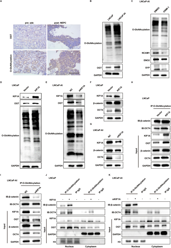
Fig. 5. KIF1A regulates activity of OGT to promote intranuclear O-GlcNAcylation of OCT4 and β-catenin.
A Representative images showing immunohistochemistry staining for O-GlcNAcylation and OGT in ade and NEPC. Scar bar = 100 μm. B Western blot of O-GlcNAcylation and OGT in LNCaP/LNCaP-AI cells. C O-GlcNAcylation and NE marker expression in LNCaP-AI cells measured by Western blot assays. LNCaP-AI cells were treated with 20 μM OSMI-1 or DMSO for 72 h. D, E Overall protein O-GlcNAcylation level in LNCaP/LNCaP-AI cells with KIF1A perturbation assessed by western blot assay. F, G Immunobloting analysis of indicated PCa cells showing changed in β-catenin and OCT4 protein level. H, I Detection of O-GlcNAcylated β-catenin and OCT4 protein in indicated PCa cells with overexpression or knockdown of KIF1A. J, K Subcellular detection of O-GlcNAcylated β-catenin and OCT4 in indicated PCa cells with overexpression/knockdown of KIF1A. ade, adenocarcinoma.

