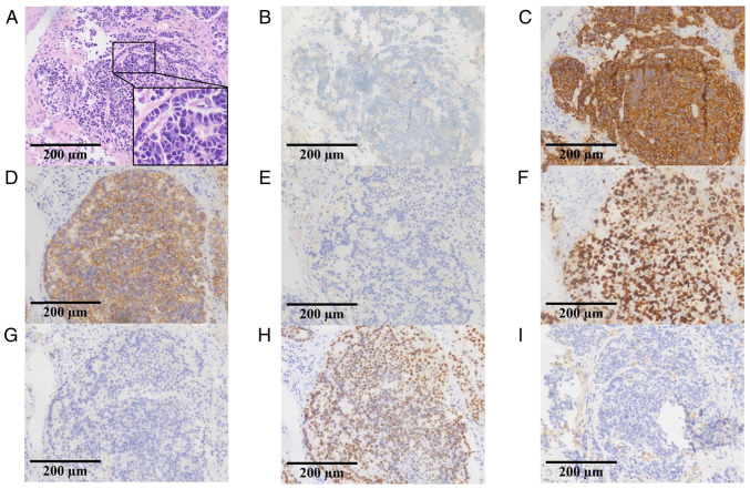
Figure 1.
Pathology and immunohistochemistry results of the biopsy specimen. (A) Hematoxylin and eosin staining revealed the presence of large cell neuroendocrine carcinoma. Immunohistochemistry results for (B) chromogranin A, (C) synaptophysin, (D) CD56, (E) NapsinA, (F) Ki-67, (G) P40, (H) thyroid transcription factor-1 and (I) programmed cell death-ligand 1 staining.

