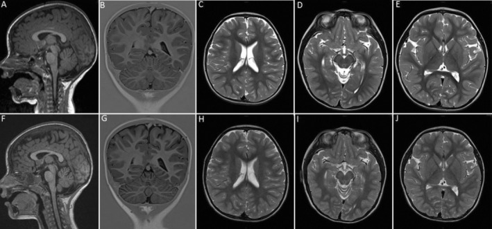
Figure 1.

Brain MRI at 2.4 years of age shows mild atrophy of the superior cerebellar vermis (A), while the cerebellar hemispheres, white matter, mesencephalon and basal ganglia are normal (B–E). Follow‐up brain MRI at 3.4 years reveals progression of the cerebellar atrophy to involve the hemispheres (F, G), while the white matter, mesencephalon and basal ganglia are still normal (H–J).
