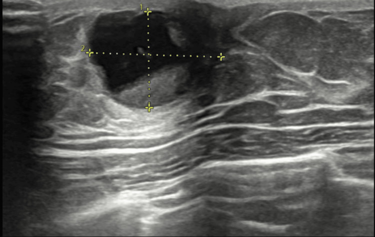
Figure 1. Ultrasonography breast done as a part of the triple assessment.
The well-defined oval-shaped mixed solid and cystic lesion measured 15 x 13 mm and was located at the 12 o'clock position of the left breast. The lesion shows a solid component adherent to the wall measuring approximately 10x11mm. Imaging features were consistent with a suspicious breast lesion. No other lesions in the left breast were seen. Suspicious-appearing lymph nodes were noted in the left axilla with maintained fatty hilum.

