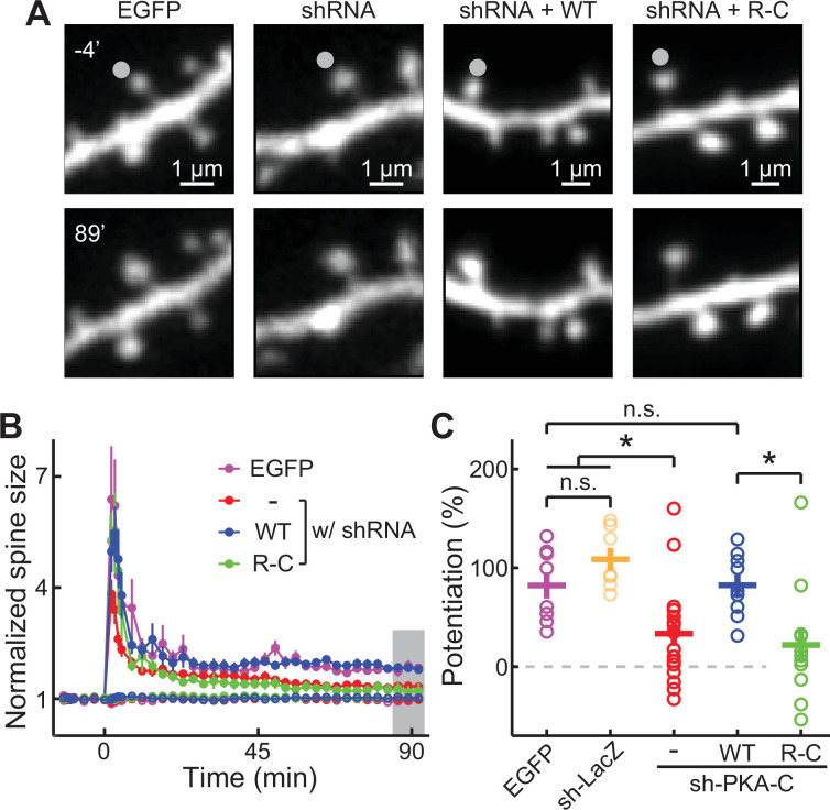
Figure 3. PKA regulation of synaptic plasticity cannot be sustained by an inseparable PKA variant.
(A–C) Representative image (A), time course (B), and the degree of potentiation (C) at the indicated timepoints in panel B of single-spine LTP experiments as triggered by focal glutamate uncaging at the marked spines (gray dot). In panel B, both stimulated spines (solid circles) and non-stimulated control spines (open circles) are shown. As in panel C from left to right, n (spines, each from a different neuron)=8, 7, 17, 11, 9. Error bars represent s.e.m.

