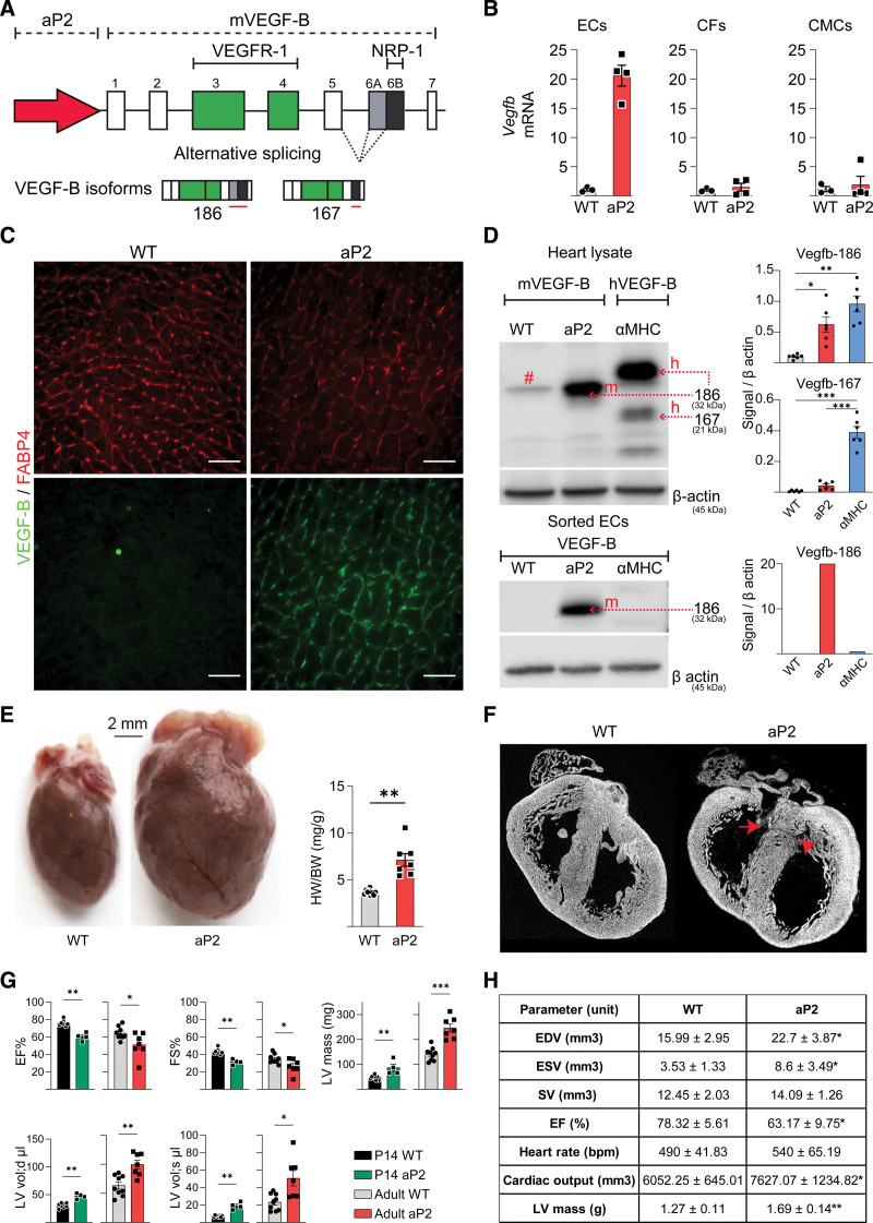
Figure 1.
Septal defects and massive pathological cardiac hypertrophy caused by autocrine VEGF-B (vascular endothelial growth factor B) expression in coronary endothelial cells (ECs). A, Schematic illustration of the aP2 (adipocyte protein 2)-VEGF-B transgene encoding both VEGF-B isoforms (VEGF-B186 and VEGF-B167). B, Quantitative real-time polymerase chain reaction (RT-qPCR) analysis of mVegfb RNA in ECs, cardiac fibroblasts (CFs), and cardiomyocytes (CMCs) isolated from hearts of 10-week-old aP2-mVEGF-B mice (n=3 wild type [WT], 4 aP2). *Unpaired Mann-Whitney t test. C, Representative immunohistochemical stainings of aP2 (FABP4 [fatty acid binding protein 4]) and VEGF-B in cryosections from the hearts of adult aP2-VEGF-B mice and their WT littermate mice. Scale bars, 50 µm. D, Western blot (WB) analysis of VEGF-B and β-actin in heart lysates (n=6) and isolated ECs from adult aP2-VEGF-B, myosin heavy chain alpha (αMHC)-VEGF-B and WT mice. Quantifications show the fold expression normalized to β-actin. # indicates a background band. *Brown-Forsythe and Welch ANOVA test with Dunnett correction. E, Macroscopic images showing the cardiac hypertrophy phenotype in aP2-VEGF-B mice and quantification of the heart weight (HW)/body weight (BW) ratio (n=9 WT, 7 aP2). Scale bar, 2 mm. *Unpaired 2-tailed t test with Welch correction. F, Ex vivo micro-computed tomography (μCT) scans of P0 aP2-VEGF-B and WT littermate hearts. Red arrows point to the septal defects. G, Echocardiography parameters from aP2-VEGF-B and WT littermate hearts at P14 (n=8 WT, 4 aP2) and at 10 wk (n=9 WT, 7 aP2). *P14 unpaired Mann-Whitney t test; adults unpaired 2-tailed t test with Welch correction. H, Cardiac magnetic resonance imaging (MRI) parameters from aP2-VEGF-B and WT littermate hearts (n=5). Values are represented as means±SD. *Unpaired Mann-Whitney t test. EDV indicates end-diastolic volume; EF, ejection fraction; ESV, end-systolic volume; LV, left ventricle; and SV, stroke volume.

