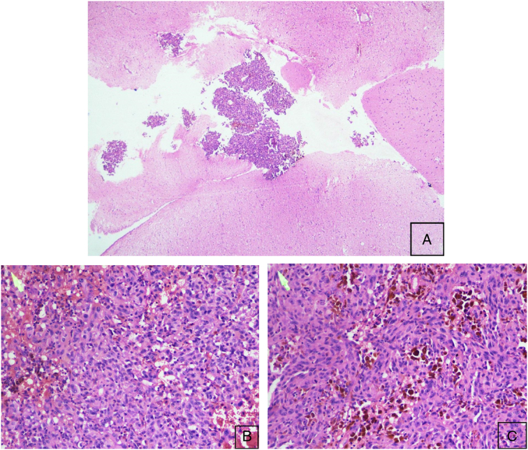
Figure 2.

Histopathological examination of malignant metastatic melanoma. (A) Microscopic examination of mass revealing the glial tissues infiltrated by the sheets of tumor cells. (Hematoxylin and eosin, HE 4×). (B, C) Discohesive round to polygonal to spindle-shaped tumor cells. Large pleomorphic nuclei with prominent nucleoli and moderate cytoplasm. Multinucleated giant cells and areas of necrosis, hemorrhages, and blood vessels (Hematoxylin and eosin, HE 20×).
