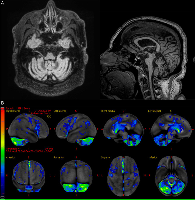
Figure. MRI and [18F] Fluorodeoxyglucose (FDG)-PET Imaging.
Research 3T volumetric MRI scan demonstrating pontine-cerebellar atrophy (A). [18F] fluorodeoxyglucose (FDG)-PET demonstrating bilateral motor/premotor, bilateral cerebellar, and striatal hypometabolism, most prominent in the posterior fossa (B).

