. 2024 Oct 29;107(5):252–263. doi: 10.4174/astr.2024.107.5.252

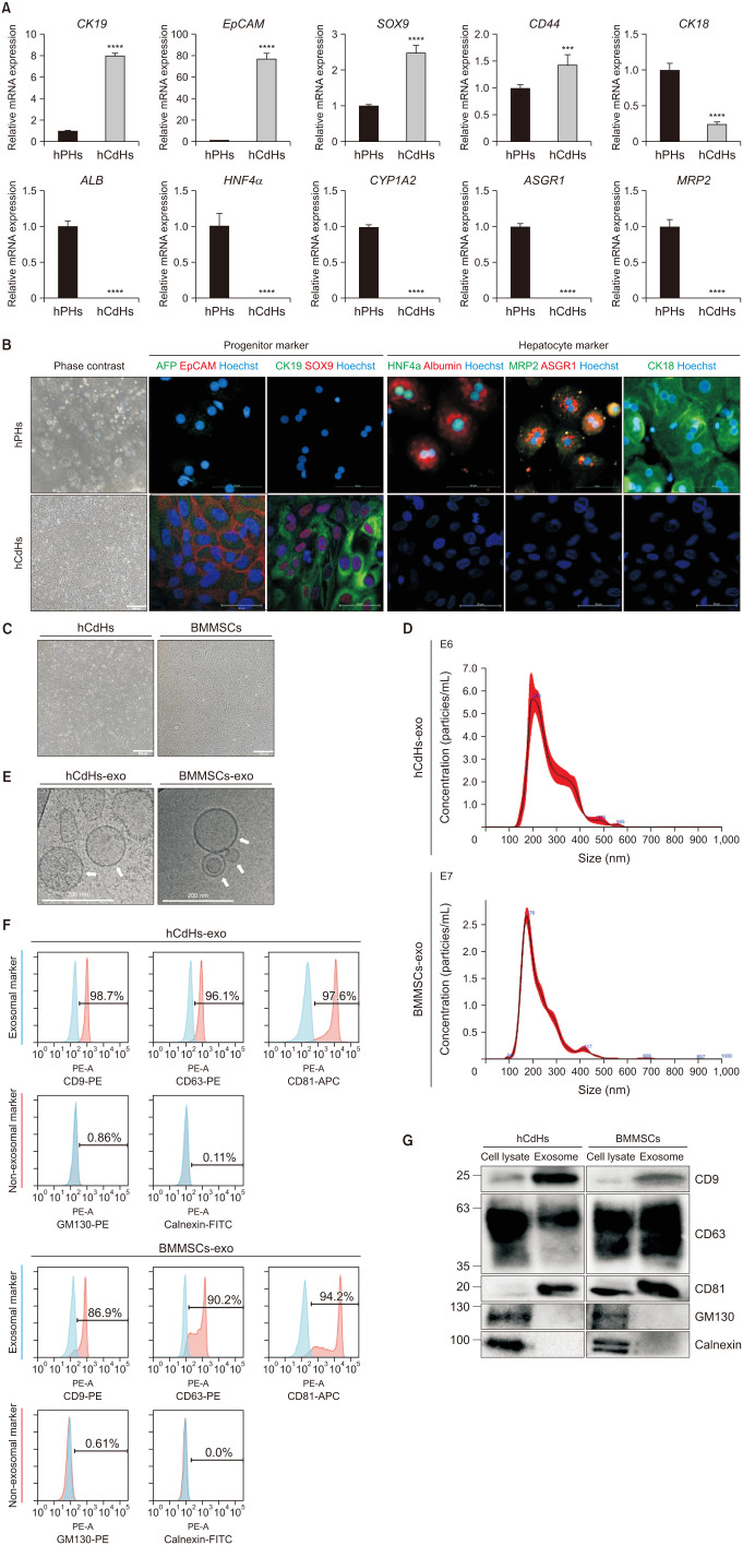
Fig. 1. Isolation and characterization of exosomes derived from human chemically derived hepatic progenitors (hCdHs) and bone marrow mesenchymal stem cells (BMMSCs). (A) Quantitative real-time PCR analysis of hepatic progenitor marker (CK19, EpCAM, SOX9, and CD44) and hepatocyte marker (CK18, ALB, HNF4α, CYP1A2, ASGR1, and MRP2) genes. GAPDH was used as an internal control. Data are shown as mean ± standard deviation (n = 3). Data were analyzed by 2-tailed t-tests (***P < 0.001, ****P < 0.0001). (B) Representative phase contrast (left) and double immunofluorescence (right) images of staining for the hepatic progenitor markers AFP (green)/EpCAM (red) and CK19 (green)/SOX9 (red), and for the hepatocyte markers HNF4α (green)/albumin (red), MRP2 (green)/ASGR1 (red), and CK18 (green). Nuclei were counterstained with Hoechst 33342 (blue). Scale bars, 500 µm and 100 µm. (C) Brightfield imaging of hCdHs and BMMSCs. Scale bars, 500 µm. (D) Size distribution of hCdHs-derived exosomes (hCdHs-exo) and BMMSCs-derived exosomes (BMMSCs-exo) as assessed by nanoparticle tracking analysis. (E) Morphological analysis of isolated hCdHs-exo and BMMSCs-exo by cryogenic transmission electron microscopy (white arrows). Scale bar, 200 nm. (F) Expression of exosomal (CD9, CD63, and CD81) and non-exosomal (GM130 and calnexin) markers was evaluated in isolated exosome samples by flow cytometry using marker-specific antibodies. (G) Expression of exosome markers in cell lysates and exosomes were confirmed by western blotting. GAPDH, glyceraldehyde-3-phosphate dehydrogenase.

Fig. 1