. 2024 Oct 29;107(5):252–263. doi: 10.4174/astr.2024.107.5.252

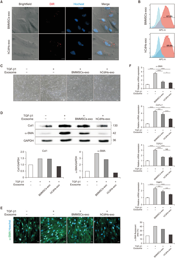
Fig. 2. Human chemically derived hepatic progenitors-derived exosomes (hCdHs-exo) suppress hepatic stellate cell activation by transforming growth factor beta 1 (TGF-β1). (A) Confocal microscopy data were acquired after processing DiR-labeled exosomes in human hepatic stellate cells (HHSteCs). Scale bars, 10 µm. (B) Flow cytometry analysis of HHSteC treated with DiR-labeled exosomes. (C) Representative phase contrast images of HHSteCs treated with bone marrow mesenchymal stem cells-derived exosomes (BMMSCs-exo) or hCdHs-exo for 48 hours in the presence of TGF-β1 (2 ng/mL). Scale bars, 100 µm. (D) Western blot analysis of HHSteC incubated with BMMSCs-exo or hCdHs-exo for 48 hours in the presence of TGF-β1 (2 ng/mL). (E) Representative immunofluorescence images of alpha smooth muscle actin (α-SMA; green). Nuclei were counterstained with Hoechst 33342 (blue). Scale bars, 100 µm. (F) Quantitative real-time PCR analysis of liver fibrosis marker genes (α-SMA, Col1α1, TGFβ1, and TIMP1). GAPDH was used as an internal control. Data are shown as the mean ± standard error of mean (n = 3 independent experiments). Data were analyzed by 2-tailed t-tests (*P < 0.05, ***P < 0.001, and ****P < 0.0001). NS, not significant.

Fig. 2