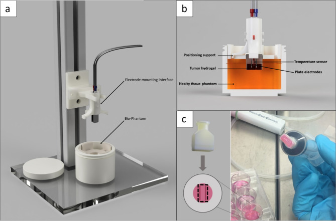
Fig. 1.
A: Experimental setup for an EP procedure including the electrode mounting interface positioned on a rail and the Bio-Phantom; B: Cross-section of the electrode mounting interface including the positioning support, temperature sensor and plate electrodes, and the Bio-Phantom consisting of the tumor hydrogel, healthy tissue phantom C: 3D-printed biopsy punch for isolating the ablation area of a hydrogel. The printed part is connected with a luer-lock system to a 20 ml syringe.

