Fig. 5.
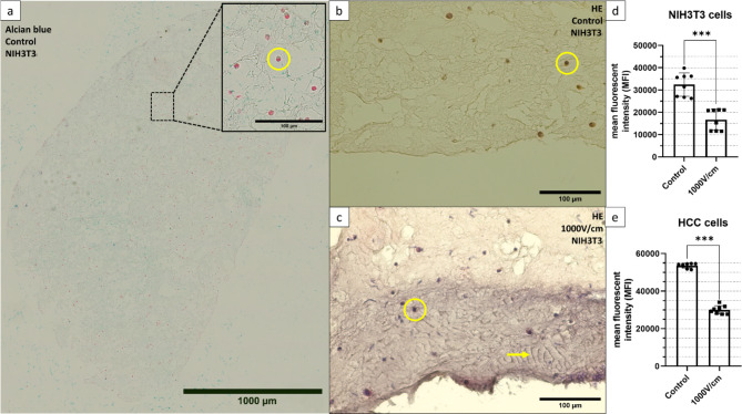
A-C Stainings of paraffin-embedded collagen-based hydrogel containing NIH3T3 cells (yellow circle). A: Alcian blue staining displaying the cell distribution throughout the section. B: HE staining of untreated control with homogenic staining of the ECM. C: HE staining of hydrogel treated with Stimulate IRE at E = 1000 V/cm with 25 cycles of 8 pulses, displaying a ~150µm wide purple-stained area at the insertion point of the plate electrode and fiber-like structures marked with a yellow arrow. D-E: Relative metabolic activity of [N=8] collagen-based hydrogels 24 hours after treatment with the Stimulate IRE at E = 1000 V/cm with 25 cycles of 8 pulses for D: Cultivated with 1 × 10⁶ HCC-827 cells. E: Cultivated with 1 × 10⁶ NIH3T3 cells. The reference were each N = 8 untreated hydrogels. Values are generated with an AlamarBlue assay with 20 hours of incubation time.

