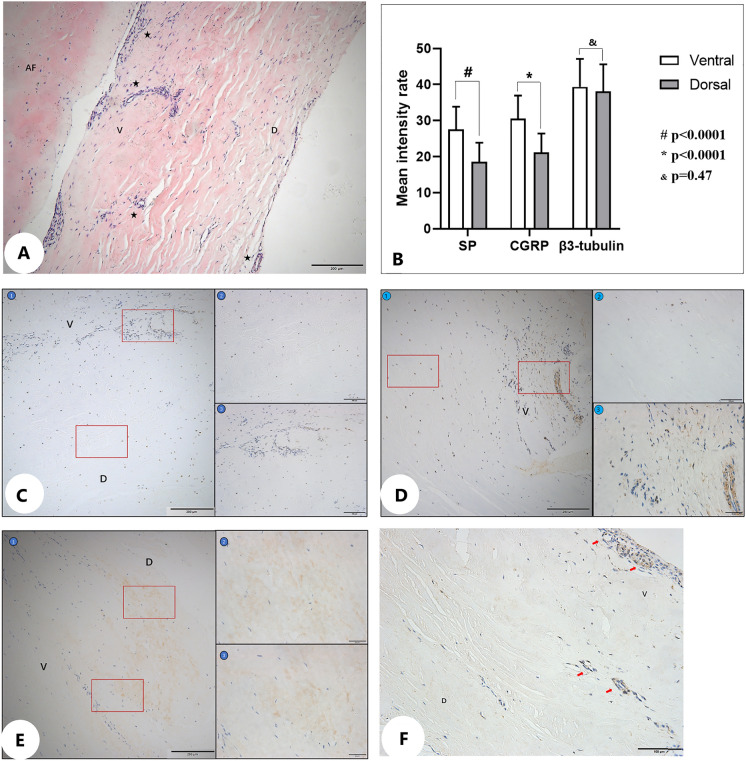
Fig. 6.
A HE staining of the PLL (V: ventral side of PLL; D: dorsal side of PLL; AF: annulus fibrosus; capillaries: ★). B Schematic diagram of positive immunoreactivity of different neuronal markers in the ventral and dorsal sides of the PLL. C Schematic diagram of positive immunohistochemical reaction of SP in the ventral and dorsal sides of the PLL (①: at ×10 objective; ②: dorsal side at ×40 objective; ③: ventral side at ×40 objective). D Schematic diagram of positive immunohistochemical reaction of CGRP in the ventral and dorsal sides of the PLL (①: at ×10 objective; ②: dorsal side at ×40 objective; ③: ventral side at ×40 objective). E Schematic diagram of positive immunohistochemical reaction of β3-tubulin in the ventral and dorsal sides of the PLL (①: at ×10 objective; ②: dorsal side at ×40 objective; ③: ventral side with ×40 objective). F Capillaries (arrows) in the PLL

