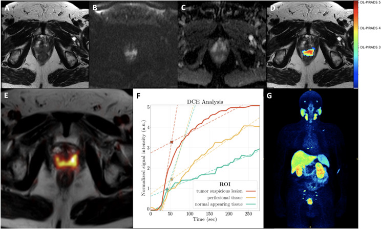
Fig. 3.
A 70-year-old patient with an initial diagnosis of PC (Gleason score 9) referred for initial staging with PET-MRI. His PSA level at the time of examination was 63.0 ng/ml. MRI showed a lesion suspicious for PC mainly in the posteromedial/-lateral peripheral zone with infiltration in the seminal vesicles (not shown) with T2w hypointensity (A) and diffusion restriction with increase in high b-value (B) and decrease in ADC map (C). The AI probability score is in good agreement with the MRI findings and also shows a high probability score for PC (DL-PIRADS 4, D). Quantitative data of DCE based on manual segmentations showed a steeper wash-in slope for tumor-suspicious lesion (TSL, red) compared to perilesional tissue (PLT, yellow) and normal appearing tissue (NAT, green). The corresponding fitted maximum (*, intersection of wash-in and wash-out slope) was 3.3 for TSL, 1.4 for PLT and 0.9 for NAT, enabling a clear distinction between TSL, PLT and NAT. All three curves showed an increasing curve pattern (F). Whole-body [18F]-PSMA-1007 PET imaging revealed the primary tumor as well as multiple PSMA-avid iliac lymph node metastases and osseous metastases in the sacral bone, thoracic spine and left scapula (G)

