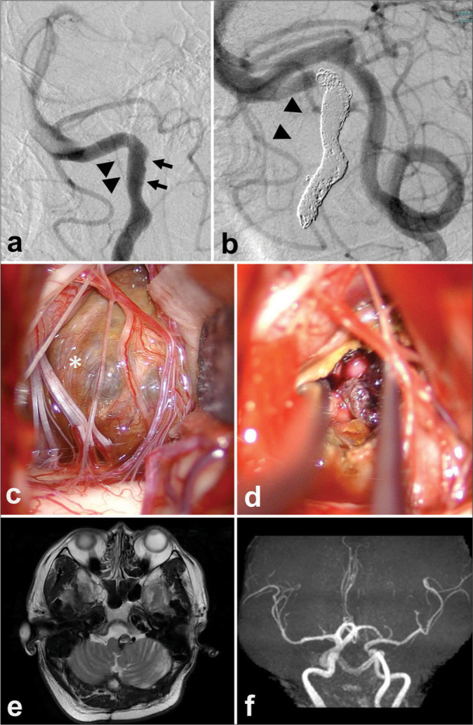
Figure 2:

Short-term two-stage operation for a patient with a thrombosed giant aneurysm of the right vertebral artery (VA). Endovascular parent artery occlusion of the affected artery (a and b) and decompressive partial removal of the giant thrombosed aneurysm through the suboccipital supracondylar fossa approach (c and d) and magnetic resonance imaging 1 year after the operation (e and f). (a)The right vertebral angiogram before the treatment showed an irregular arterial wall (arrow) that coincided with a giant aneurysm that is not described by internal thrombosis. An anterior spinal artery (ASA) originating from the V4 portion of the right VA is shown (arrowheads). (b)The left vertebral angiogram after the treatment shows complete obliteration of the right VA by coil embolization just before the origin of the ASA (arrowheads). (c)The intraoperative photograph shows the thrombosed aneurysm wall (asterisk). (d) Internal decompression of the thrombosed aneurysm. (e) T2WI 1 year after the surgery shows that the thrombosed aneurysm is reduced in size and brainstem compression is improved. (f)The right VA is not described on magnetic resonance angiography 1 year after the operation.
