
Figure 3:
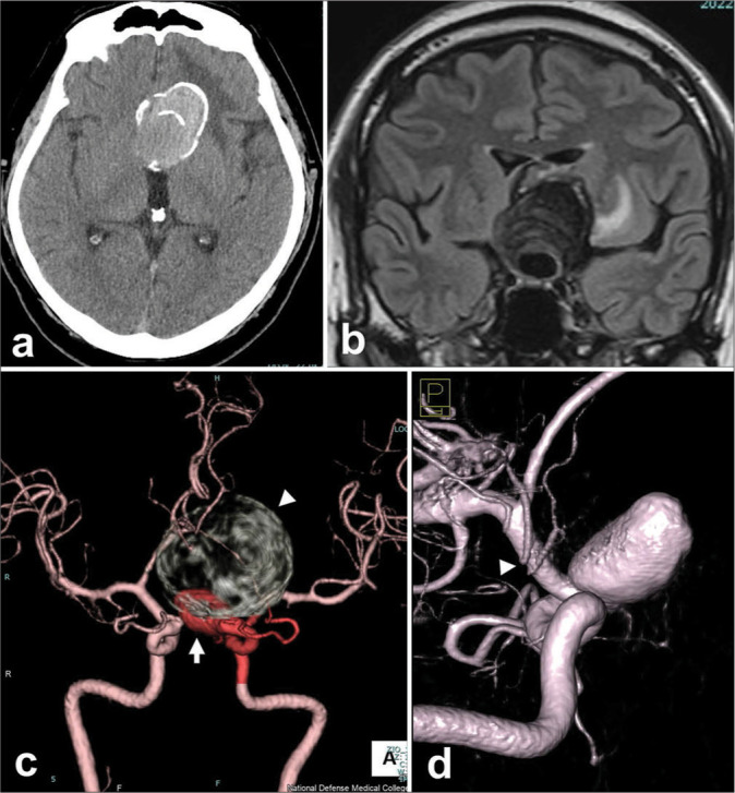
Pre-operative radiograms of a patient with a giant thrombosed aneurysm of the left internal carotid artery. (a and b) A large mass of 50 mm in diameter is shown at the left frontal region, which involves calcifications on computed tomography (CT) (a) and surrounding edema in the left frontal lobe on coronal magnetic resonance imaging of fluid-attenuated inversion recovery (b). (c)The whole configuration of the left carotid artery aneurysm, which consists of a large thrombosed and calcified part (arrowhead) and a flow-remnant part (arrow), is shown by the contrast-enhanced CT. (d)Three-dimensional rotational angiography revealed a 7.5 mm distance from the distal neck of the aneurysm to the origin of an anterior choroidal artery (arrowhead).