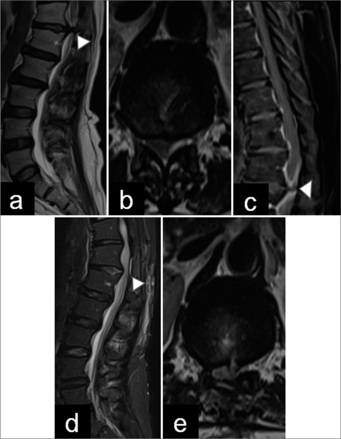
Figure 1:

Pre- and post-operative magnetic resonance imaging (MRI). (a and b) Preoperative sagittal and axial T2-weighted images demonstrate a T12–L1 right-sided disc extrusion (arrowhead) occupying more than 90% of the spinal canal. (c) Post-contrast T1 MRI shows partial enhancement around the disc herniation. (arrowhead in image c is herniated disc), (d and e) Postoperative sagittal and axial T2-weighted images show an adequate decompression of the spinal canal. Postoperative changes in the laminectomy and discectomy are observed (arrowhead in image d is soft tissue inflamation).
