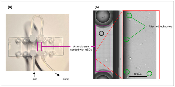
Figure A1.
(a) Channel slides seeded with lung ECs (luECs in vivo) were perfused with 1 × 10⁶ fluorescently labelled, activated leukocytes under unidirectional flow conditions for 30 min at a flow rate of 1 dyn/cm2. The complete channel is marked with a purple square. (b) View of one analyzed area (marked in red) of a channel (purple) at a magnification of 10×. Adherent leukocytes (green) were determined by automatic particle counting using ImageJ software in all areas of the channel (purple). Attached leukocytes (marked with green circles) were identified by color (green), size (above 5 pixels), and round shape (0.5 to 1.0).

