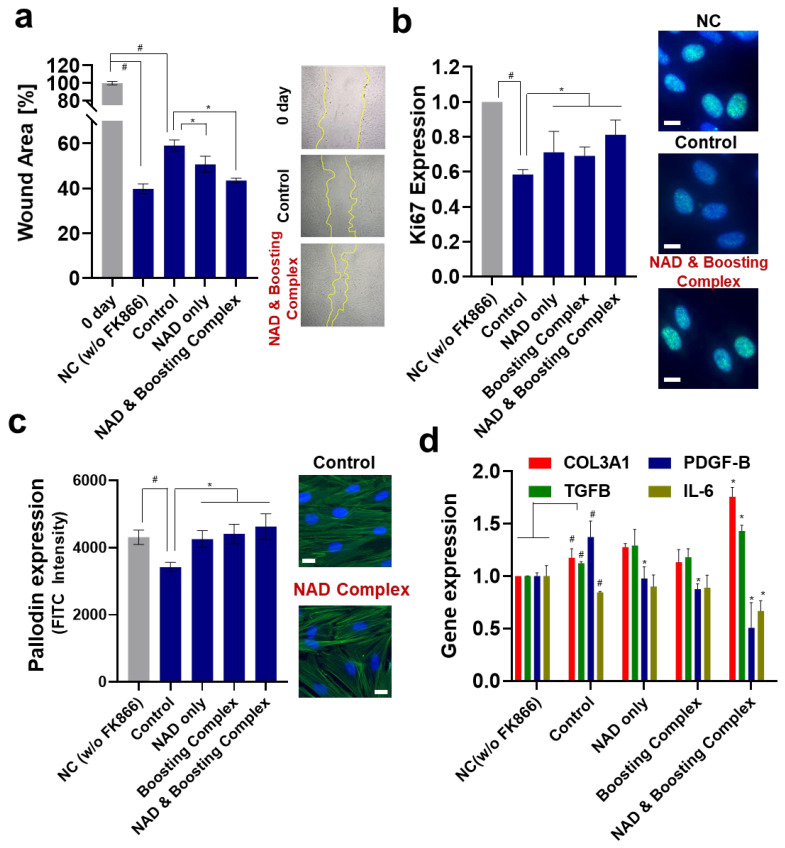
Figure 4.
Elevation of NAD+ enhances wound healing of fibroblasts. (a) Wound scratch assay: the wound area was measured after 24 h. (b) Ki67 expression: the FITC field intensity (ex/em = 488/520) was measured and normalized against the nuclear staining intensity (Hoechst 33342). Scale bar (White) = 20 m. (c) Actin/phallodin staining: similar to Ki67, FITC intensity was measured and normalized using nuclear staining intensity (Hoechst 33342). Scale bar (White) = 20 m. (d) RT-qPCR was performed to assess wound-healing-related gene expression. All experiments were performed in triplicate. Student’s t-test was performed to determine the significance between negative control and control group (# Significantly different results (p < 0.05)). One-way ANOVA (Dunnett’s test) was performed for comparison between control and experimental groups (* Significantly different results (p < 0.05)).

