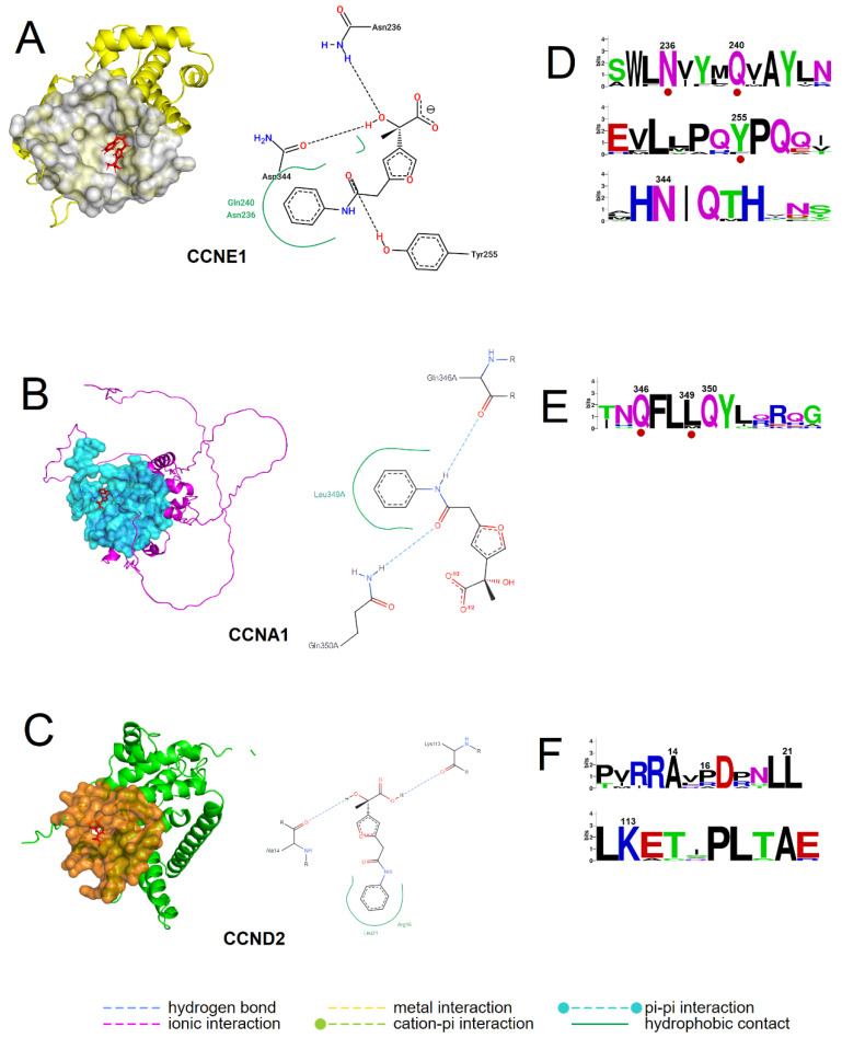
Figure 5.
Identification of amino acids involved in the interaction of the novel ligand with cyclin E1, A1, and D2. (A–C) Docking poses of the ligand against CCNE1, CCNA1, and CCND2. Left: Cartoon representation of target proteins and stick representation of the ligand compound. Right: 2D view of the protein–ligand binding residues. (D–F) Logos of corresponding conserved cyclin sequence motifs. The numbers denote the positions of the amino acids that are involved in the ligand–cyclin interaction. The residues reported to reside in the cyclin–CDK interface are indicated by dark red dots. The height of each letter is proportional to the frequency of the occurrence of the corresponding amino acid at that position.

