Figure 6.
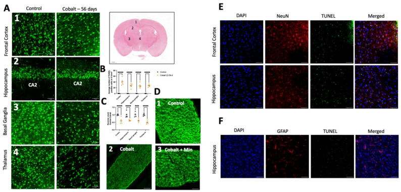
Immunostaining and quantification of neuronal loss and cell death. (A) Neurons were labeled with NeuN antibody in the frontal cortex, hippocampus, thalamus, and basal ganglia. (B) Quantification of the mean number of neurons in these brain areas indicated neuronal loss in the cobalt-treated group. (C) Quantification of TUNEL+ cells/area in the frontal cortex, hippocampus, thalamus, and basal ganglia. **** p < 0.0001, *** p < 0.001, Student t-test. Number of mice: n = 5 in each group. Data shown are mean ± SEM. Scale bar = 50 µm. LD = low dose. (D1) Illustration of normal myelin in a control mouse. (D2) Extensive demyelination in a cobalt-treated mouse. (D3) Partial preservation of myelin in a minocycline-treated mouse on MBP immunohistochemical staining. Scale bar: 50 µm. (E) TUNEL and neuronal marker NeuN+ staining after daily IP treatment for 56 days yielded positive findings. (F) GFAP and TUNEL did not overlap, indicating that astrocytes were less susceptible to cobalt toxicity.

