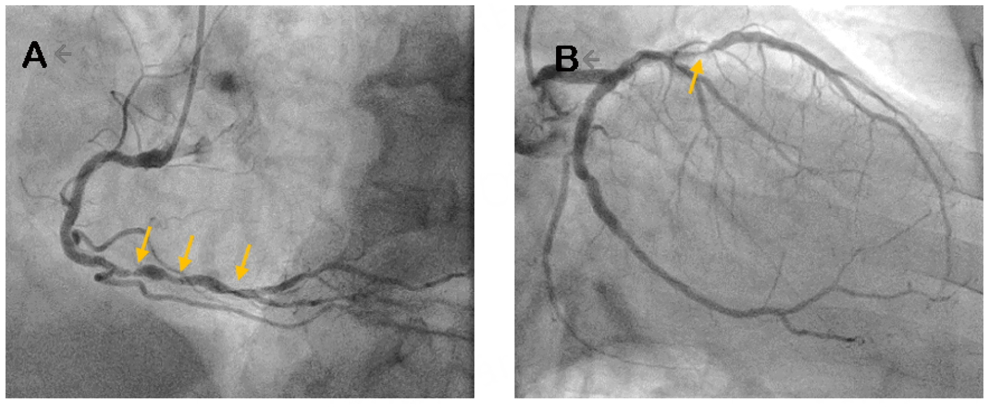
Figure 3.

Coronary angiogram images of a 75-year-old man with a history of STEMI and PCI to RCA, LAD, and LCF who presented with angina and was discussed by a Heart Team (Patient #2). (A) representative image of the right coronary artery showing several areas of distal stenoses (yellow arrows). (B) A representative image of the left coronary arteries and in-stent restenosis of the LAD (yellow arrow). STEMI: ST elevation myocardial infarction; PCI: percutaneous intervention; RCA: right coronary artery; LAD: left anterior descending; LCF: left circumflex.
