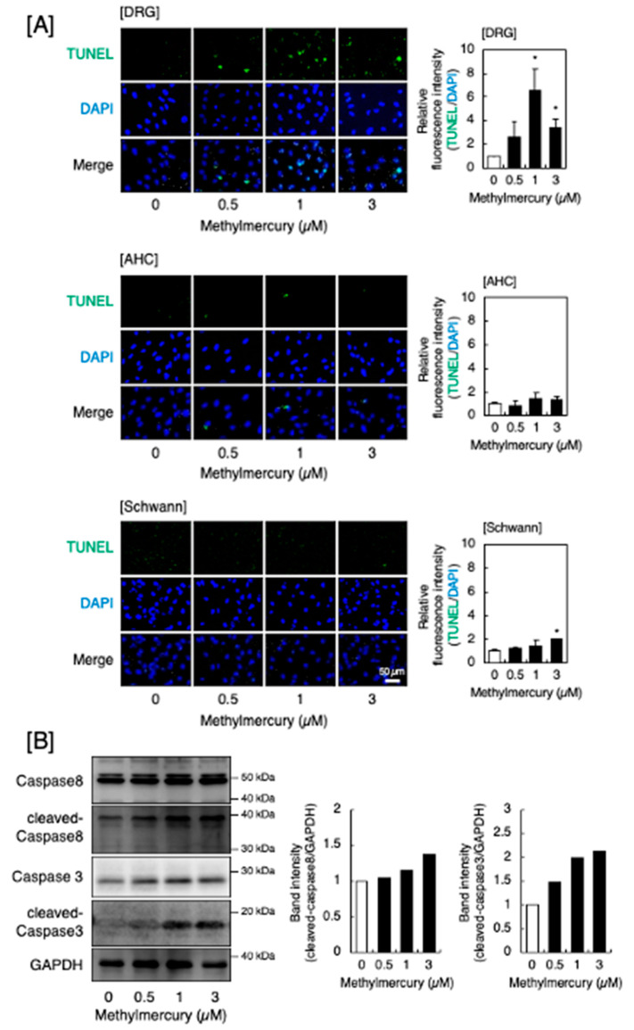
Figure 2.
Apoptosis induced by methylmercury. (A) Representative images of the TUNEL assay (left panels, 100× magnification) and the quantitative analysis (right panels) of cultured DRG neurons (upper panels), AHCs (middle panels), and Schwann cells (lower panels) after treatment with methylmercury (0.5, 1, and 3 µM) for 24 h. Values are means ± S.E. of three technical replicates. Significantly different from the corresponding control, * p < 0.05. (B) Activation of caspase 8 and caspase 3 by methylmercury in DRG neurons. The cells were treated with methylmercury (0.5, 1, and 3 µM) for 24 h, and the expression of caspase 8, cleaved caspase 8, caspase 3, and cleaved caspase 3 was determined by Western blot analysis. Glyceraldehyde-3-phosphate dehydrogenase (GAPDH) was used as an internal control. The representative Western blot of at least two experiments is shown.

