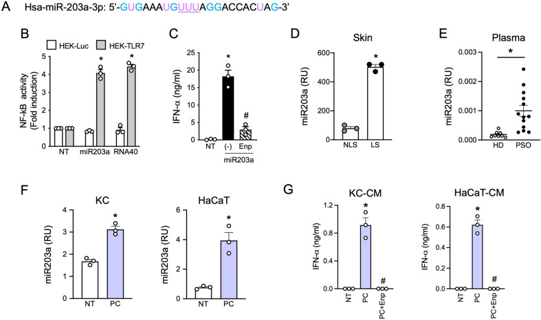
Fig. 1.
miR203a is upregulated in psoriatic patients and released by inflamed keratinocytes. (A) Sequence of synthetic hsa-miR203a-3p. G and U are highlighted in light blue and purple respectively while the TLR7 full binding motif “UUU” is underlined. (B) HEK293 cells stably transfected with a NF-kB reporter gene and human TLR7 or luciferase alone were stimulated with 10 µg/ml of miR203a or RNA40 for 24 h. NF-κB activation was evaluated in terms of luciferase activity (fold of induction) over unstimulated cells (NT). Data are expressed as mean ± SEM (n = 3); *P < 0.05 vs. “NT” by 1-way ANOVA with Dunnett’s post hoc test. (C) pDCs were pretreated or not with Enpatoran (Enp, 1 µM) for 1 h and then stimulated with miR203a for 24 h. IFN-α secretion was evaluated by ELISA (mean ± SEM, n = 3); *P < 0.05 vs. “NT” or #P < 0.05 vs. (-) by paired Student’s t test. (D) miR203a expression from paired non lesional (NL) and lesional (L) skin biopsies from 3 psoriatic patients was investigated by qPCR. Data are expressed as mean ± SEM of Relative Units (RU) compared to cel-miR39; *P < 0.05 vs. “NLS” by paired Student’s t test. (E) miR203a expression in the plasma of healthy donors (HD, n = 8) and psoriatic patients (PSO, n = 13) by qPCR; *P < 0.05 vs. “HD” by Mann-Whitney test. (F) Primary keratinocytes (KC) and HaCaT cells were stimulated with a mix of psoriatic cytokines (PC, each cytokine at 50 ng/ml) for 24 h and the expression of miR203a in the supernatant was assessed by qPCR (mean ± SEM, n = 3); *P < 0.05 vs. “NT” by paired Student’s t test. (G) pDCs were pretreated or not with Enpatoran for 1 h and then stimulated with conditioned media of KC or HaCaT (CM, 50% vol/vol). IFN-α production was evaluated by ELISA (mean ± SEM, n = 3); *P < 0.05 vs. “NT” or #P < 0.05 vs. (-) by paired Student’s t test

