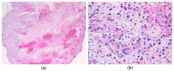
Figure 1.
Hematoxylin and eosin staining of the biopsy tissue showing a myxoid and necrotic tumor with pleomorphic tumor cells, giant cells, and partial rhabdoid features that were classified as undifferentiated embryonal sarcoma of the liver (a) 12.5× magnification; (b) 400× magnification. Immunohistochemistry revealed positivity for CD56, but negativity for SMA, desmin, pancytokeratin, CD99, myogenin, MyoD1, MDM2, S100, EMA, WT1, and CD34 in the tumor cells. The Ki67 proliferation index was measured up to 40% in the sample.

