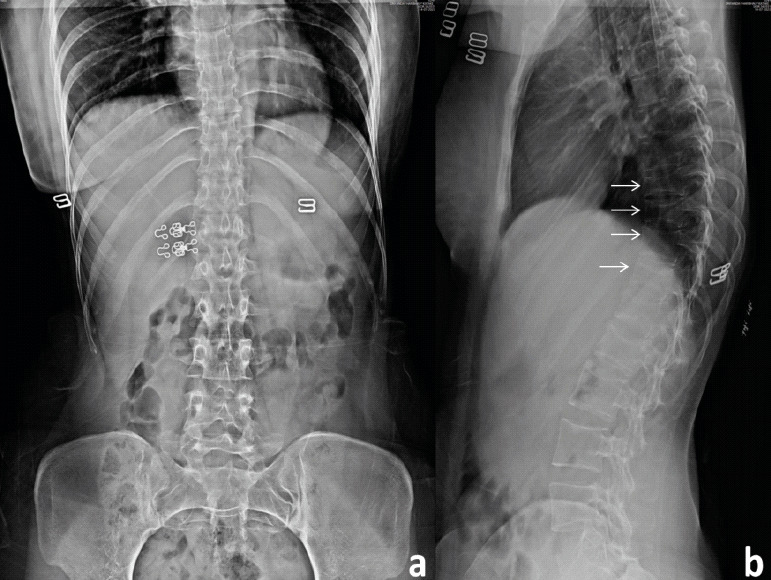
Figure 1.

X-ray: Anteroposterior (a) and lateral view (b) of the dorsolumbar spine: showing a loss of height from D9-12 vertebral bodies along with decreased cortical thickness and loss of bony trabeculae associated with smooth biconcave deformities, squared-off depressions of the end-plates combined with compressions from the adjacent discs which were representative of cod fish vertebrae suggestive of multiple osteoporotic vertebral fractures.
