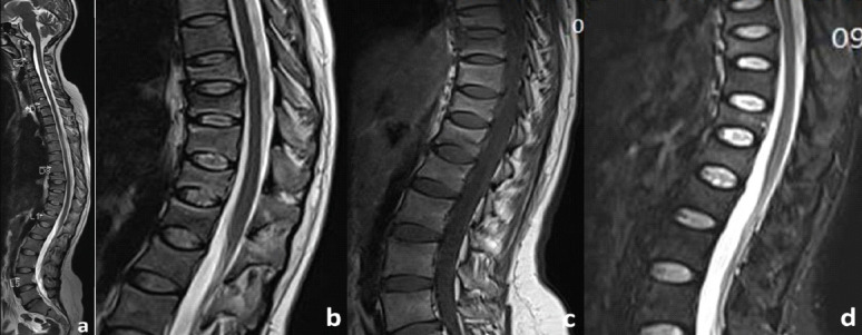
Figure 2.

MRI: Sagittal images of whole spine (a) T2 showing anterior wedge collapse at D9-D12 vertebra. Sagittal images of dorsolumbar spine T2 (b) hyperintense, T1 (c) hypointense, and STIR (d) hyperintense signal in the D9 and D10 vertebra with normal signal intensity in the intervening disk spaces with preservation of intraosseous fat.
