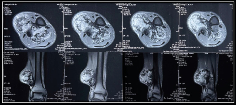
Figure 4.

MRI Right leg denoting pedunculated, large, chondroid lesion arising from the lateral cortex of the distal tibia with adjacent cortical thickening giving rise to a large lobulated mass lesion mixed with osseous and chondroid signals (7.3 × 5.7 × 4.4 cm, AP × TR × CC). The lesion bulged in the anterior and posterior intermuscular plane of the leg causing significant compression and displacement of the extensor and flexor muscle compartments. Chronic pressure changes over the medial cortex of the distal fibula with mild pressure-related periostitis on the fibular cortex with no signs of invasion of the fibula.
