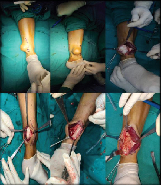
Figure 6.

Intraoperative outlines of the lesion. The margins of the surface were identified and marked with a skin marker to delineate the extent of the incision. After appropriate dissection, the lesion was exposed and excised. The tumor was resected out from the tibial cortex and the normal cortical surface was visualized. The excised tumor mass was sent for histopathological diagnosis. The fibular surface was exposed and was evident for compression effects of the lesion showing periosteal irritation, though the bone was stable and required no fixation.
