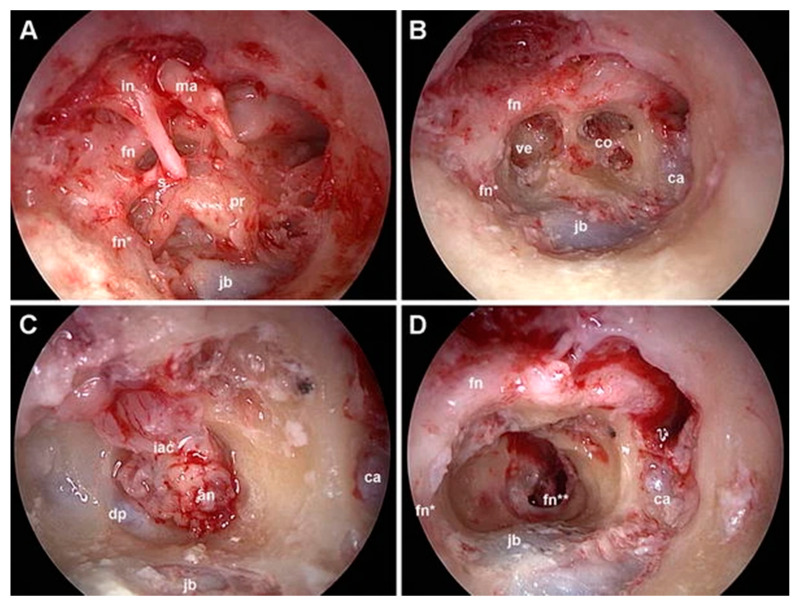
Figure 4.
Exclusive endoscopic transcanal transpromontorial approach, right ear. (A): wide drilling of the external auditory canal is performed, thus exposing the tympanic cavity. (B): after ossicular chain removal and promontory drilling, the turns of the cochlea and vestibule are exposed. (C): the dissection proceeds, reaching the IAC; the dura is opened, showing in this case a vestibular schwannoma. (D): final tympanic cavity after acoustic neuroma removal with preserved facial nerve. In, incus; ma, malleus; s, stapes; ve, vestibule; ca, carotid artery; jb, jugular bulb; co, cochlea; fn, facial nerve (tympanic tract); fn*, facial nerve (mastoid tract); fn**, facial nerve (IAC segment); iac, internal auditory canal; an, acoustic neuroma; dp, reflection of the dura from the porus.

