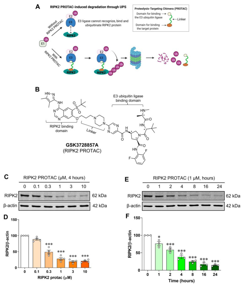
Figure 1.
Dose- and time-dependent degradation of RIPK2 by its proteolysis-targeting chimera in SIM-A9 cells. Model of RIPK2 degradation mediated by its proteolysis-targeting chimera (PROTAC) molecule GSK3728857A (A) and the molecular structure of the RIPK2 PROTAC (B). Representative immunoblots and graphs showing degradation of RIPK2 by RIPK2 PROTAC in microglial cells. (C,D) Incubation with various concentrations of RIPK2 PROTAC (0–10 μM) for 4 h degrades RIPK2 in a dose-dependent manner. (E,F) Similarly, time-dependent degradation of RIPK2 by RIPK2 PROTAC (1 μM) was also observed. One-way ANOVA with Bonferroni post-test; * p < 0.05 and *** p < 0.001 compared with control conditions. Data are normalized to β-actin and represented as mean ± SEM from three to four independent experiments.

