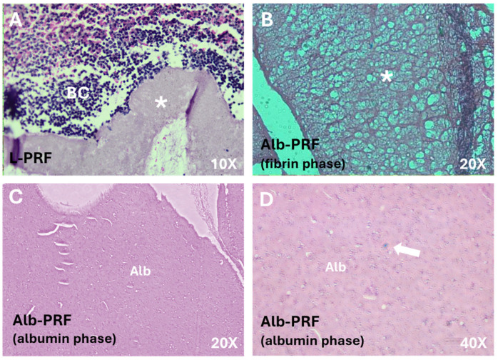
Figure 1.
Images of histological sections of the membranes. (A) L-PRF membrane at its extremity, where both a dense fibrin network (*) and the buffy coat (BC) can be identified with a high density of cells; Alb-PRF forms a biphasic material with a fibrin network phase shown in (B), connected to a dense albumin barrier (Alb) shown in (C). This albumin portion is also cellularized (arrow), as it can be seen at a higher magnification (D).

