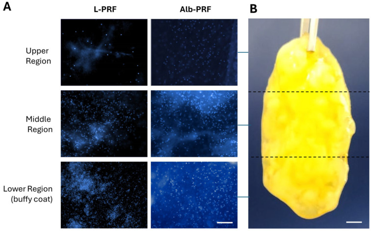
Figure 3.
Qualitative panel of cellular distribution in the structure of L-PRF and Alb-PRF membranes (A). Images were taken from three different portions of the membranes, as shown for an Alb-PRF membrane with approximately 3 cm of length (B). Cell nuclei were evidenced by fluorescence microscopy after staining with DAPI in the upper, middle, and lower regions of the L-PRF membrane and visualization of three random fields of the Alb-PRF membrane (images obtained with a ×20 objective). The scale bar in (A) represents 100 μm, while in (B) it represents 3 mm.

