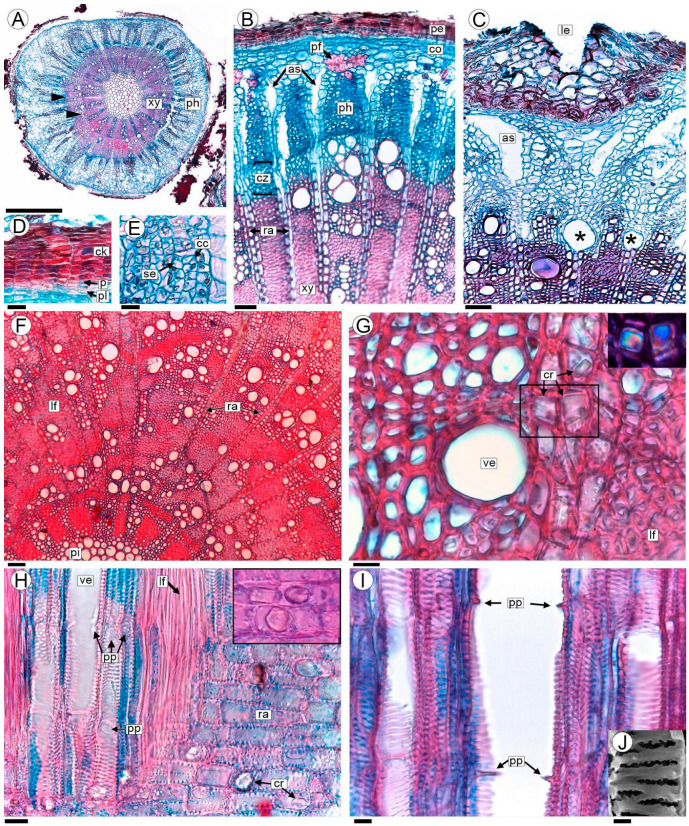
Figure 2.
Anatomy of the non-parasitized stem of A. trijuga in transversal (A–G), and radial longitudinal sections (H,I) are analyzed with a light microscope (LM). (A) Transection of 2-year-old non-parasitized stem; (B,C) phloem and cambial zone in a stem with vessels in the process of formation (*); (D) periderm; (E) secondary phloem; (F) wood anatomy showing its diffuse porosity; (G) detail of vessels, paratracheal parenchyma and libriform fibers; the crystals in the area indicated with a square are shown in the inset (polarized light); (H) vessels, libriform fibers and rays; crystals in the inset; (I) vessels showing simple perforation plates; (J) detail of vestured pits. Abbreviations: as: air spaces; cc: companion cells; ck: cork; co: cortex; cr: prismatic crystals; cz: cambial zone; lf: libriform fibers; le: lenticels; p: phellogen; pe: periderm; pf: primary phloem fibers; ph: secondary phloem; pi: pith; pl: phelloderm; pp: simple perforation plate; ra: rays; se: sieve elements; ve: vessels; xy: xylem. Scales: (A) 0.5 mm; (B,C,F) 50 µm; (D,H) 20 µm; (E,G,I) 10 µm; (J) 2 µm.

