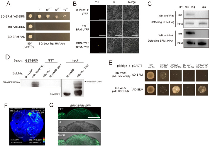
Fig 3. BRM interacts with DRN mediating WUS-DRN-BRM complex.
(A) Y2H shows the interaction of DRN-BRM. The combinations with BD and AD empty vectors were introduced as negative controls. Yeast cells in a series of dilutions were grown on the selective medium (SD/−Leu/−Trp/−His/−Ade). Two independent experiments were performed with similar results. (B) BiFC exhibiting the interaction of DRN-BRM in tobacco leaves. YFP was split into N-terminus and C-terminus, which were fused to DRN and BRM, respectively. BF, bright field. Scale bars, 100 μm. Two independent experiments were performed with similar results, and 5 leaves were analyzed for each group. (C) Co-IP showing the interaction of DRN and BRM in Arabidopsis. 35S::DRN-Flag and 35S::BRM-3×HA were transformed into Arabidopsis protoplasts. The anti-Flag antibody was used for IP. Instead of the anti-Flag antibody, IgG was used for IP as the negative control. Two independent experiments were performed with similar results. (D) Pull-down assay showing the interaction of DRN and BRM directly. Recombinant proteins were expressed in E. coli and the anti-His antibody was used for immunoblot analysis. Two independent experiments were performed with similar results. (E) Y3H results showing that WUS fails to directly bind to BRM (upper lane) and that introducing DRN produces the WUS-DRN-BRM complex (bottom lane). SD/−Leu/−Trp/−His and SD/−Leu/−Trp/−His/−Ade were used for selective media. Lacking Met in addition allowed the transcription of pMET25 promoter to produce DRN in the bottom lane but not in the upper lane. Two independent experiments were performed with similar results. (F) Split-luciferase complementation assays were used to test the association between WUS and BRM mediated by DRN in tobacco leaves. Two independent experiments were performed with similar results. Six leaves were analyzed for each independent replicate. (G) BRM::BRM-GFP lines were used to detect the distribution of BRM-GFP in SAMs. Green, GFP signal; BF, bright field. Scale bars, 50 μm. Eight apices were analyzed. BiFC, bimolecular fluorescence complementation; SAM, shoot apical meristem; Y2H, yeast two-hybrid; Y3H, yeast three-hybrid.

