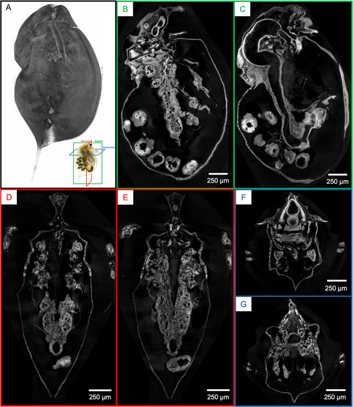
Fig 1. Whole-organism imaging of PTA-stained D. magna at cell resolution enables histology-like cross sections.
(A) 3D volume rendering shows the scanning electron microscopy-like surface rendering. Sagittal (B, C), coronal (D, E), and transverse (F and G) cross-sections can be obtained from one sample after imaging. B to G represent 5 μm thick micro-CT slabs.

