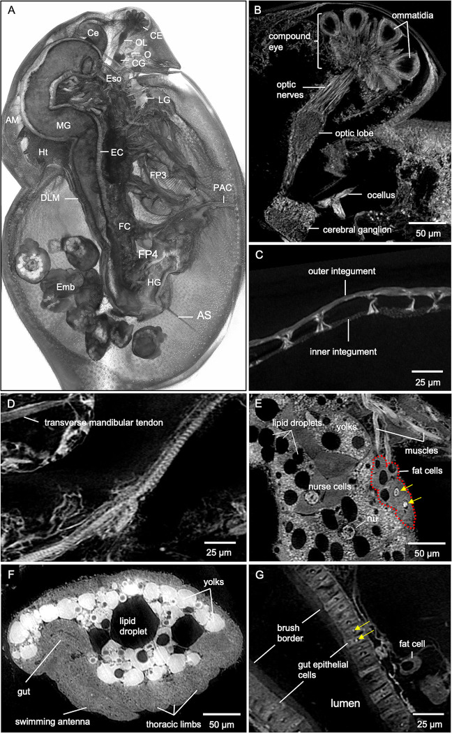
Fig 2. Microanatomic features of an adult female D. magna from synchrotron-based micro-CT imaging at 0.5 μm per pixel resolution.
(A) 3D rendering at the mid-section of the sagittal plane with various organs, organ substructures, and cell types indicated. AM, antennal muscles; AS, abdominal setae; Ce, hepatic ceca; CE, compound eye; CG, cerebral ganglia; DLM, dorsal longitudinal muscles; EC, gut epithelial cells; Emb, developing embryos; Eso, esophagus; FC, fat cells; FP3, filter plates on third pair of thoracic limbs; FP4, filter plates on fourth pair of thoracic limbs; HG, hindgut; Ht, heart; LG, labral glands; MG, midgut; O, ocellus, OL, optic lobe; PAC, post-abdomen claws. Highlights of microanatomical features include (B) ommatidia of the compound eye, optic nerves, optic lobe, cerebral ganglia, and ocellus. Other features include (C) filamentous actin bundle pillars between inner and outer carapace integuments and (D) muscle striations along the thoracic muscle. (E) Details in the ovary include nucleus (nu) of the oocyte, nurse cells, yolks, and lipid droplets. Nucleoli (yellow arrows) in fat cells are also clearly visible. (F) Recognizable details in the developing embryo include precursors of the gut, swimming antennae, and thoracic limbs. (G) The brush border and epithelial cells with nucleoli (yellow arrows) within the nuclei are visible in the gut. B represents a 5 μm thick micro-CT slab; C represents a 5μm thick micro-CT slab; D to G represent individual 0.5 μm thick micro-CT slices.

