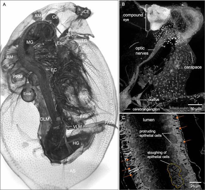
Fig 3. Microanatomic features of a wild-type female D. magna with an atypical eye.
(A) 3D rendering at the mid-section of the sagittal plane with various organs and organ substructures indicated. AM, antennal muscles; AS, abdominal setae; Ce, hepatic ceca; CE, compound eye; CG, cerebral ganglia; DLM, dorsal longitudinal muscles; EC, gut epithelial cells; Emb, developing embryos; Eso, esophagus; FP3, filter plates on third pair of thoracic limbs; HG, hindgut; Ht, heart; L, labrum; LG, labral glands; MG, midgut; O, ocellus; PAC, post-abdomen claws. (B) Micro-CT provides details where the compound eye (CE) has less than 22 ommatidia with irregular shape and arrangement. The optic nerves (ON) connect directly to the cerebral ganglia without an optic lobe. (C) Whole organism micro-CT imaging also revealed abnormalities in the gut, with an excessive number of epithelial gaps (orange arrows), protruding and sloughing of the gut epithelial cells. B and C represent 25 and 5 μm thick micro-CT slabs, respectively.

