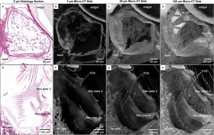
Fig 4. Visualization of anatomical structures using micro-CT slabs of various thicknesses.
The 5 μm thick micro-CT slab of heart and filter plates (B and F, respectively) resembles the 5 μm thick histological tissue section (A and E, respectively). Thicker micro-CT slabs (50 μm) allow visualization of (C) more heart wall muscles and (G) overlapping long setae of the filter plates. Micro-CT slabs of 100 μm showed (D) the posterior rotator muscles of the mandible (PRM) and fat cells around the heart, (H) both filter-cleaning spines (FCS) on the second pair of thoracic limbs, and both filter plates on the third pair of thoracic limbs (H). DLM, dorsal longitudinal muscles.

