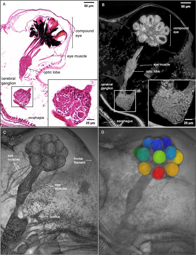
Fig 5. Histology section, micro-CT image, and 3D Rendering of D. magna vision system.
Comparison of D. magna visual system using 5 μm thick histology section and micro-CT slab of the same thickness (A and B, respectively). Insets show details of cerebral ganglion where the micro-CT slab demonstrates the near histological resolution of micro-CT imaging at 0.5-micron resolution. (C) 3D rendering featuring the vision system allows clear visibility of the frontal filament that connects to the ocellus, the eye muscle bundles, and their insertion into the compound eye. (D) Image segmentation of structures of interest (crystalline cones shown here) allows the isolation of specific structures for measurement or quantitative analysis.

